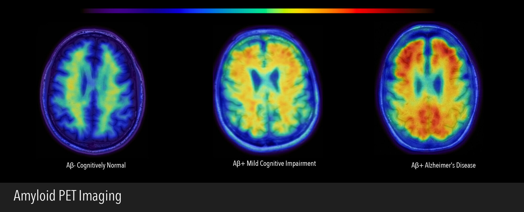
β-amyloid (Aβ) is a major constituent of the amyloid plaque and is believed to play an important role in the pathogenesis of Alzheimer’s disease. In the aging brain and Alzheimer’s disease Aβ aggregates into plaques which are deposited in the brain. The aggregated forms of Aβ appear to disrupt brain function and lead to a cascade of detrimental events. We can image Aβ in the brain using PET scans. During PET imaging, a radioactive tracer binds to amyloid plaques and allows us to image their distribution in the brain. The amount and regional distribution of Aβ in conjunction with other imaging and genetic biomarkers can be used to characterize a variety of neurodegenerative disorders including Alzheimer’s disease, vascular dementia, dementia with Lewy bodies, and frontotemporal dementia. Abnormal amyloid accumulation is also found in the brains of healthy, cognitively normal older people. How this accumulation affects the brain, and whether it is a sign of future problems, is a major focus of research in the lab. The multiple techniques that we use in the lab including PET scanning, MRI, and cognitive testing allow us to investigate the very beginnings of Alzheimer’s disease and to understand the transition between normal cognitive aging and Alzheimer’s disease.